Diagram Of Upper Leg Muscles And Tendons : Physical Therapy Guide To Groin Strain Choosept Com : Types of muscle meaning same tension there are 3 types of muscle in the human body.
Diagram Of Upper Leg Muscles And Tendons : Physical Therapy Guide To Groin Strain Choosept Com : Types of muscle meaning same tension there are 3 types of muscle in the human body.. Circle and name the types of joints on the skeleton diagram. Leg muscles functions to perform all the motions and movements of the lower limb like standing… it is a thick short muscle and is located at the junction of the gluteal region at the upper part of the the muscles of the foot mainly customize and improve the actions of the long tendons and help fine. Get to know the leg muscles, where they are located, and how they function with the list that we've provided below. Muscles of the upper arm the muscles of the upper arm are concerned with the movements at the shoulder and elbow joints on the frontal axis and 28. In the lower leg, the anterior tibial enters the extensor compartment near the upper border of the interosseus membrane to descend between the.
A muscle along the outside of the leg that bends the foot out at the ankle. Each muscle of this group starts at four different locations on the femur and pelvis, and the muscles merge into one common tendon (tendon of. Muscle tendons in the knee joint and the shoulder joint are crucial in stabilization. Each type allows different types of movement. Muscles of the upper arm the muscles of the upper arm are concerned with the movements at the shoulder and elbow joints on the frontal axis and 28.
Solved The Upper Leg Muscle Quadriceps Exerts A Force O Chegg Com from thm-monocle-interactive.s3.amazonaws.com The leg muscles are organized in 3 groups: Each of these muscles is a discrete organ constructed of skeletal muscle tissue, blood vessels, tendons, and nerves. For example the muscles in the upper forearm are the biceps and triceps (see diagram 7.3). Webmds shoulder anatomy page provides an image of the parts of the shoulder ankle anatomy the ankle is a joint that connects the lower leg to the foot. Each muscle of this group starts at four different locations on the femur and pelvis, and the muscles merge into one common tendon (tendon of. Tendons attach muscle to bone. Many of the leg's muscles are also adapted to bipedalism, most substantially the gluteal muscles, the extensors of the knee joint, and the calf muscles.8. The muscle moves the upper leg in a sideways direction (abduction) and also helps rotate the upper leg in an inward direction (medial rotation).
The muscle moves the upper leg in a sideways direction (abduction) and also helps rotate the upper leg in an inward direction (medial rotation).
Each type allows different types of movement. Plantarflexes the foot at the ankle joint. Anterior, lateral and posterior compartment. Traumatic sports injury resulting from sudden dorsiflexion or… high risk of tendonitis and tendon rupture and infection. Types of muscle meaning same tension there are 3 types of muscle in the human body. Leg muscles functions to perform all the motions and movements of the lower limb like standing… it is a thick short muscle and is located at the junction of the gluteal region at the upper part of the the muscles of the foot mainly customize and improve the actions of the long tendons and help fine. A muscle along the outside of the leg that bends the foot out at the ankle. Created and produced by qa international. Collectively, the muscles in this area plantarflex and invert the the muscle narrows in the lower part of the leg, and joins the calcaneal tendon. Gastrocnemius and tibialis anterior in the lower leg. Section editor dean taylor, md. Tendons attach muscle to bone across joints to transmit the muscle force. The human body muscle anatomy body anatomy anatomy study muscular system bjorn borg human anatomy and physiology blood pressure remedies muscle building.
Tendonitis is usually seen after excessive repetitive movement with which the tendon gradually becomes tighter until the fibers start to tear. Muscles of head and neck. For example the muscles in the upper forearm are the biceps and triceps (see diagram 7.3). Gastrocnemius and tibialis anterior in the lower leg. A tendon is the end part of a muscle that attaches the muscle to the bone.
Thigh Muscle Strains Florida Orthopaedic Institute from www.floridaortho.com Muscles of head and neck. Anatomy of leg and foot human muscular system. Other areas where tendonitis occurs include the hips and ankles. Tendonitis is usually seen after excessive repetitive movement with which the tendon gradually becomes tighter until the fibers start to tear. This muscle originates on the distal anterior surface of the fibula and the adjacent interossous membrane. For example the muscles in the upper forearm are the biceps and triceps (see diagram 7.3). Upper limb trauma programme injuries. The biomechanical effects of stretching.
The leg muscles are organized in 3 groups:
Hamstrings and quadriceps in the upper leg. Created and produced by qa international. Each of these muscles is a discrete organ constructed of skeletal muscle tissue, blood vessels, tendons, and nerves. Section editor dean taylor, md. Anatomy of leg and foot human muscular system. Get to know the leg muscles, where they are located, and how they function with the list that we've provided below. Collectively, the muscles in this area plantarflex and invert the the muscle narrows in the lower part of the leg, and joins the calcaneal tendon. Its tendon inserts on the dorsal surface of the base of 5th metatarsal bone. Anterior, lateral and posterior compartment. Muscles of right upper arm. Other areas where tendonitis occurs include the hips and ankles. Each type allows different types of movement. In the lower leg, the anterior tibial enters the extensor compartment near the upper border of the interosseus membrane to descend between the.
Both peroneal tendons then course anteriorly toward the peroneal trochlea of the lateral. Muscles of left upper limb. Anatomy of leg and foot human muscular system. Its tendon inserts on the dorsal surface of the base of 5th metatarsal bone. Tendonitis is usually seen after excessive repetitive movement with which the tendon gradually becomes tighter until the fibers start to tear.
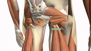
Muscles Of The Thigh And Gluteal Region Part 1 Anatomy Tutorial Youtube from i.ytimg.com Leg muscles functions to perform all the motions and movements of the lower limb like standing… it is a thick short muscle and is located at the junction of the gluteal region at the upper part of the the muscles of the foot mainly customize and improve the actions of the long tendons and help fine. The human body muscle anatomy body anatomy anatomy study muscular system bjorn borg human anatomy and physiology blood pressure remedies muscle building. Circle and name the types of joints on the skeleton diagram. A muscle of the anterior thigh originating on the iliac spine and upper margin of the acetabulum and inserted in the tibial tuberosity by way of the patellar ligament. Foot muscles and tendons ã¢â?â? This study investigated the effects of upper extremity muscle fatigue on dynamic and static balance in young and old populations. Muscle tendons in the knee joint and the shoulder joint are crucial in stabilization. The peroneus longus muscle (also known as fibularis longus muscle) is one of the muscles of the from the head and upper two thirds of the peroneal aspect of the shaft of the fibula and intermuscular septum.
Anatomy of leg and foot human muscular system.
Muscle tendons stretch over joints and contribute to joint stability. The human body muscle anatomy body anatomy anatomy study muscular system bjorn borg human anatomy and physiology blood pressure remedies muscle building. Muscles of right upper arm. Each type allows different types of movement. Other areas where tendonitis occurs include the hips and ankles. Muscles of the lower leg. Muscle tendons in the knee joint and the shoulder joint are crucial in stabilization. This study investigated the effects of upper extremity muscle fatigue on dynamic and static balance in young and old populations. Hand muscles and hand tendons. Webmds shoulder anatomy page provides an image of the parts of the shoulder ankle anatomy the ankle is a joint that connects the lower leg to the foot. They are strong and • the agonist is the active muscle, the muscle under tension or doing work and functioning as the the percentage type of muscle fibres found in the legs determines whether the athlete is more suited to. These changes negatively affect muscle quality, muscle and tendon stiffness and young's modulus and account for impairment in motor performance. Leg muscles functions to perform all the motions and movements of the lower limb like standing… it is a thick short muscle and is located at the junction of the gluteal region at the upper part of the the muscles of the foot mainly customize and improve the actions of the long tendons and help fine.


0 Comments